Penfield Homunculus: Characteristics And Functions
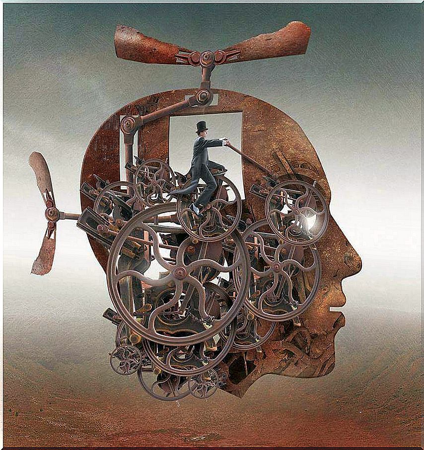
Our brain is extraordinary. We have been studying it for years and we have not yet discovered all its possibilities. It is like the universe, infinite and full of surprises. Perhaps for this reason, when new functions or brain areas are discovered, an attempt is made to simplify the finding. That’s what happened to the well-known Penfield Homunculus.
The Penfield Homunculus was first described by Dr. Wilder Penfield between the 1940s and 1950s. This Canadian neurosurgeon sought to explain and cure neurological diseases such as epilepsy. Thus, one of his best known works was undoubtedly that of neurostimulation.
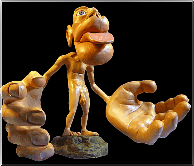
By applying small and controlled shocks, something very interesting was discovered. In our brain there is a small area that makes up the sensory map of our body. This structure reflects the sensitivity of each of the parts of our anatomy. He decided to represent this area as if it were a human form, giving rise to the Penfield Homunculus.
What makes this representation special is to be aware that we have areas in our body that are more sensitive to stimulation than others. Thus giving rise to a deformed, disproportionate man, where the most sensitive areas show greater size than those that are less so.
Now, this is not all, shortly after the existence of a new figure. In this way, we could say each of us has two “homunculi”, one sensory and the other motor, both very different but with points in common.
Characteristics and functions of the Penfield Homunculus
Studies such as those carried out by the neurologists Di Noto P, Newman L, Wall S, Einstein in 2013, update in depth those first bases on these structures that were established between 1937 and 1954 thanks to the neurosurgeon Wilder Penfield.
It should be noted, first of all, that we should not expect to see two “human” figures embedded in our brain. Dr. Penfield outlined this similarity by seeing that each sensory area was correlated to each part of our body. For example, in these structures parts such as the hand and each of our fingers are located next to each other. Let’s see it in detail.
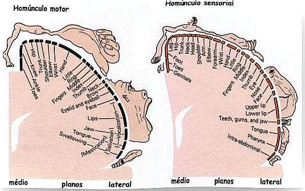
Motor homunculus or primary motor cortex:
The motor homunculus or primary motor cortex is located right next to the sensory homunculus. It is located exactly in the central sulcus of the frontal cortex. This area is the most important for the motor functioning of our body.
- Its function is to regulate and control the motor movements of our body. It does so in collaboration with other areas, such as the supplemental motor cortex and inputs received from the Thalamus.
- That is why its appearance is slightly different from that of the sensory homunculus: its mouth, eyes and especially its hands are enormous due to the greater specificity in the location of receptors and motor nerves.
A curiosity of this area is the following: it develops differently in each one of us. This implies that the speed of its development is unique and personal. It depends on which parts of the body are used the most and how they will have better motor skills or more trained in general.
Sensory homunculus or primary somesthetic cortex:
The sensory homunculus represents the primary somesthetic cortex or what is the same, the tactile, pressure or pain sensitivity of our body. It is located in the parietal lobe, just at its junction with the frontal lobe. Explained another way, the sensory homunculus comprises Broadman’s areas 1, 2, and 3.
- In this area our body diagram is represented in a contralateral way, or what is the same, in a laterally inverted way.
- This means that the right representation of our body is represented in the left area of this brain area and the left in the right part. Although it may surprise us, it is something very common in the functioning of our brain.
It should be noted that this sensory area receives most of the information projections from our body through the Thalamus.
Remember, the thalamus is the area of integration of the different sensory sources of our brain. Thanks to it, we perceive our world in an integrated way and not separately according to the sense that perceives it.
- The sensory homunculus is also in charge of our proprioception. Thanks to it, we regulate posture and our body knows the state of our organs and our muscles. And although it is strange to us, of how we are from within.
All this makes this area vital to our well-being, both physical and emotional. In fact, thanks to this structure we have that special sensitivity sensitivity in our face, our lips …
The phantom limb, the main disease of the Penfield Homunculus
The Penfield Homunculus, as we already know, collects and integrates our entire body representation, whether sensory or motor. Thus, it is interesting to know that an alteration in this area can result in a curious disease: that of the phantom limb.
- When suffering from this disease, the brain continues to feel or perceive the sensations of an amputated limb.
- This condition has been detailed by doctors, the sensory area that represents this part, continues to send the sensation of pain from our brain .
This means that even if the limb is amputated, due to the activity of the neurons of the sensory homunculus, we cannot stop feeling it. Now, it must be said that as this study explains, this discomfort usually disappears after two years.
As we can see, a discovery driven by curiosity through electrical brain stimulation has opened up a universe of possibilities. Thanks to him, we have realized the importance of each touch on our skin and of our brain and emotional development.